 sciencedrawings
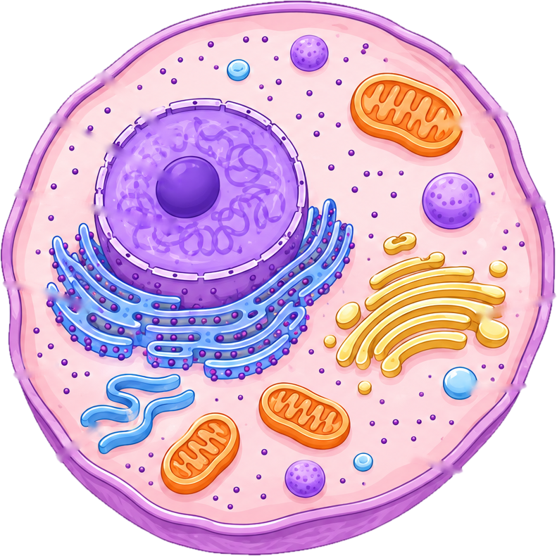 Nucleus
Nucleolus
Chromatin
Rough ER
Ribosomes
Smooth ER
Cell membrane
Mitochondrion
Lysosome
Golgi apparatus
Vesicle
Cytoplasm
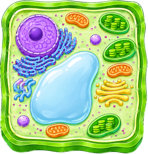
Animal Cell Diagram
A clear animal cell diagram with the main organelles labeled: nucleus, mitochondria, Golgi apparatus, endoplasmic reticulum and more. Also as a blank version for quizzes.
PNG 1400×900 · HD 2800×1800 · SVG with editable labels
Free for personal, classroom and commercial use. A credit is appreciated, not required. Paste this under it:
<a href="https://sciencedrawings.com/">Drawing by sciencedrawings.com</a>What each part does
- Nucleus
- Holds the cell’s DNA and controls what the cell does.
- Nucleolus
- A dense spot inside the nucleus where ribosomes are made.
- Chromatin
- The DNA and proteins inside the nucleus, loosely coiled when the cell is not dividing.
- Rough ER
- Folded membranes studded with ribosomes that make and transport proteins.
- Ribosomes
- Tiny structures that build proteins, floating in the cytoplasm or attached to rough ER.
- Smooth ER
- Membranes without ribosomes that make lipids and help break down toxins.
- Cell membrane
- The thin, flexible outer layer that controls what goes in and out of the cell.
- Mitochondrion
- The powerhouse of the cell: it releases energy from food by respiration.
- Lysosome
- A small sac of enzymes that breaks down waste and worn-out cell parts.
- Golgi apparatus
- Packages and ships proteins to where they are needed.
- Vesicle
- A tiny membrane bubble that carries materials around the cell.
- Cytoplasm
- The jelly-like fluid that fills the cell and holds the organelles.