 sciencedrawings
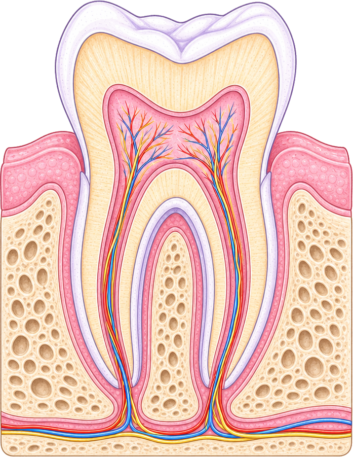 Crown
Gum
Jaw bone
Root
Blood vessels
Enamel
Dentin
Pulp
Cementum
Nerve
Tooth Diagram
A molar cut in half showing the crown, root, enamel, dentin, pulp, gum, cementum, nerve and blood vessels. Labeled and blank versions.
PNG 1400×900 · HD 2800×1800 · SVG with editable labels
Free for personal, classroom and commercial use. A credit is appreciated, not required. Paste this under it:
<a href="https://sciencedrawings.com/">Drawing by sciencedrawings.com</a>Label the tooth
A free labeling quiz: drag each label to the box it belongs in, or tap a label and then tap a box. Great for revision, homework and class.
What each part does
- Crown
- The part of the tooth above the gum that you can see and chew with.
- Gum
- The soft pink tissue that covers the jaw bone and holds the tooth in place.
- Jaw bone
- The bone the roots sit in, which anchors the tooth firmly.
- Root
- The part of the tooth below the gum that anchors it in the jaw bone.
- Blood vessels
- They carry blood into the pulp to keep the tooth alive and nourished.
- Enamel
- The hard white outer layer of the crown, the hardest substance in the body.
- Dentin
- The layer under the enamel that makes up most of the tooth and supports it.
- Pulp
- The soft centre of the tooth that holds its nerves and blood vessels.
- Cementum
- A thin bony layer covering the root that helps attach the tooth to the jaw.
- Nerve
- It senses heat, cold and pressure and sends pain signals to the brain.
More like this
 Brain Diagramlabeled + blank
 Circulatory System Diagramlabeled + blank
 Digestion Process Diagramlabeled + blank
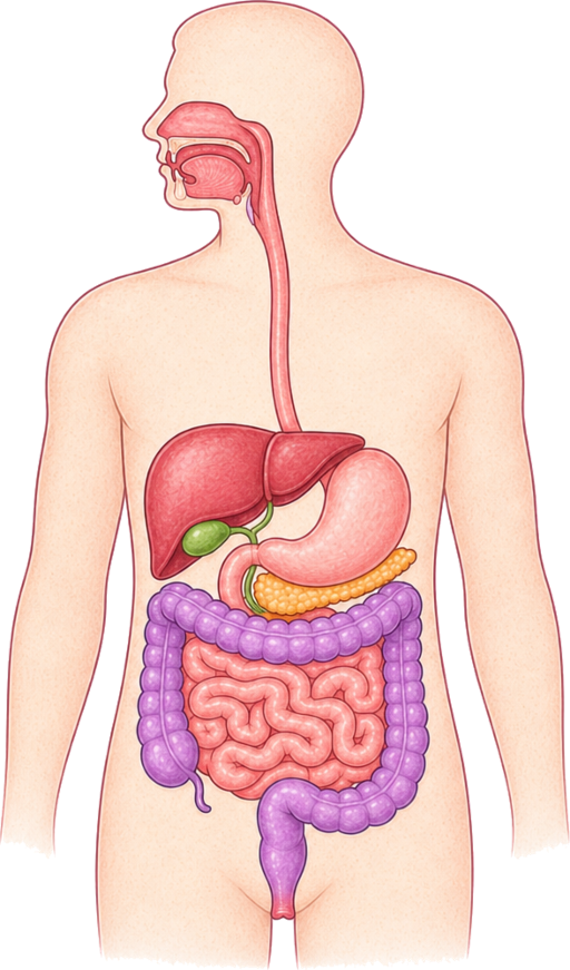 Digestive System Diagramlabeled + blank
All human body drawings · Download the human body pack (ZIP)