 sciencedrawings
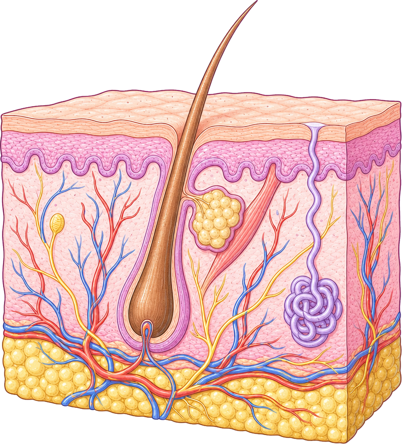 Epidermis
Dermis
Hair follicle
Blood vessels
Hypodermis
Hair
Sebaceous gland
Nerve
Sweat gland
Skin Diagram
A cross-section of human skin: epidermis, dermis and hypodermis with a hair follicle, glands, blood vessels and a nerve. Labeled and blank versions.
PNG 1400×900 · HD 2800×1800 · SVG with editable labels
Free for personal, classroom and commercial use. A credit is appreciated, not required. Paste this under it:
<a href="https://sciencedrawings.com/">Drawing by sciencedrawings.com</a>Label the skin
A free labeling quiz: drag each label to the box it belongs in, or tap a label and then tap a box. Great for revision, homework and class.
What each part does
- Epidermis
- The thin outer layer of skin that makes a waterproof barrier against germs.
- Dermis
- The thick middle layer holding blood vessels, nerves, glands and hair follicles.
- Hair follicle
- A pocket in the skin where a hair grows from its root.
- Blood vessels
- Carry blood that feeds the skin and help control body temperature.
- Hypodermis
- The deepest layer, mostly fat, which stores energy and keeps the body warm.
- Hair
- A strand of dead keratin cells that grows out of the skin and helps keep the body warm.
- Sebaceous gland
- Makes sebum, an oil that keeps the hair and skin soft and waterproof.
- Nerve
- Carries signals for touch, pain, heat and cold from the skin to the brain.
- Sweat gland
- A coiled gland that makes sweat, which cools the body as it evaporates.
More like this
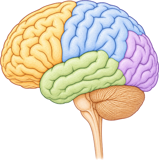 Brain Diagramlabeled + blank
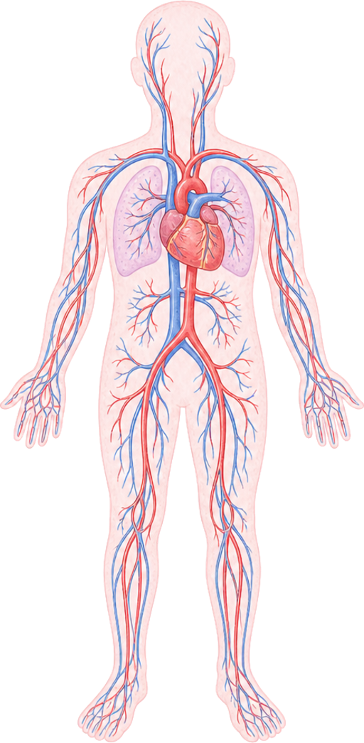 Circulatory System Diagramlabeled + blank
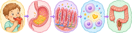 Digestion Process Diagramlabeled + blank
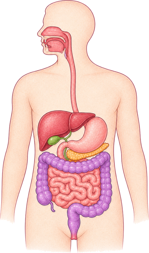 Digestive System Diagramlabeled + blank
All human body drawings · Download the human body pack (ZIP)