 sciencedrawings
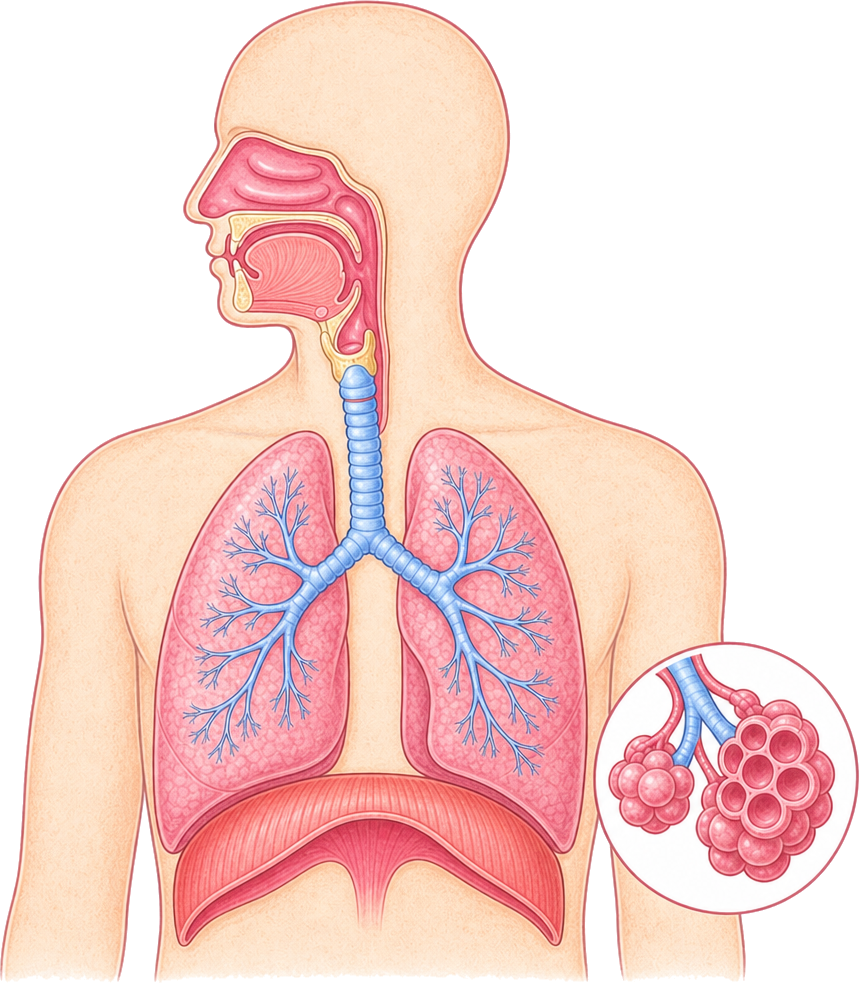 Nasal cavity
Mouth
Bronchioles
Lungs
Diaphragm
Pharynx
Larynx
Trachea
Bronchi
Alveoli
Respiratory System Diagram
The human respiratory system: nasal cavity, pharynx, larynx, trachea, bronchi, lungs, diaphragm and alveoli. Labeled and blank versions.
PNG 1400×900 · HD 2800×1800 · SVG with editable labels
Free for personal, classroom and commercial use. A credit is appreciated, not required. Paste this under it:
<a href="https://sciencedrawings.com/">Drawing by sciencedrawings.com</a>Label the respiratory system
A free labeling quiz: drag each label to the box it belongs in, or tap a label and then tap a box. Great for revision, homework and class.
What each part does
- Nasal cavity
- Air enters through the nose here, where it is warmed, moistened and filtered.
- Mouth
- A second way in for air, used when breathing hard or when the nose is blocked.
- Bronchioles
- Smaller and smaller tubes that branch from the bronchi and carry air deep into the lungs.
- Lungs
- The organs where oxygen goes into the blood and carbon dioxide comes out.
- Diaphragm
- A dome of muscle under the lungs that flattens to pull air in when you breathe.
- Pharynx
- The throat: a passage shared by air and food behind the nose and mouth.
- Larynx
- The voice box at the top of the trachea, holding the vocal cords.
- Trachea
- The windpipe: a tube held open by rings of cartilage that carries air to the lungs.
- Bronchi
- The two branches of the trachea, one leading into each lung.
- Alveoli
- Tiny air sacs with thin walls where oxygen and carbon dioxide swap with the blood.
More like this
 Brain Diagramlabeled + blank
 Circulatory System Diagramlabeled + blank
 Digestion Process Diagramlabeled + blank
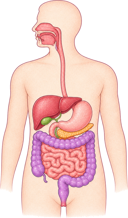 Digestive System Diagramlabeled + blank
All human body drawings · Download the human body pack (ZIP)