 sciencedrawings
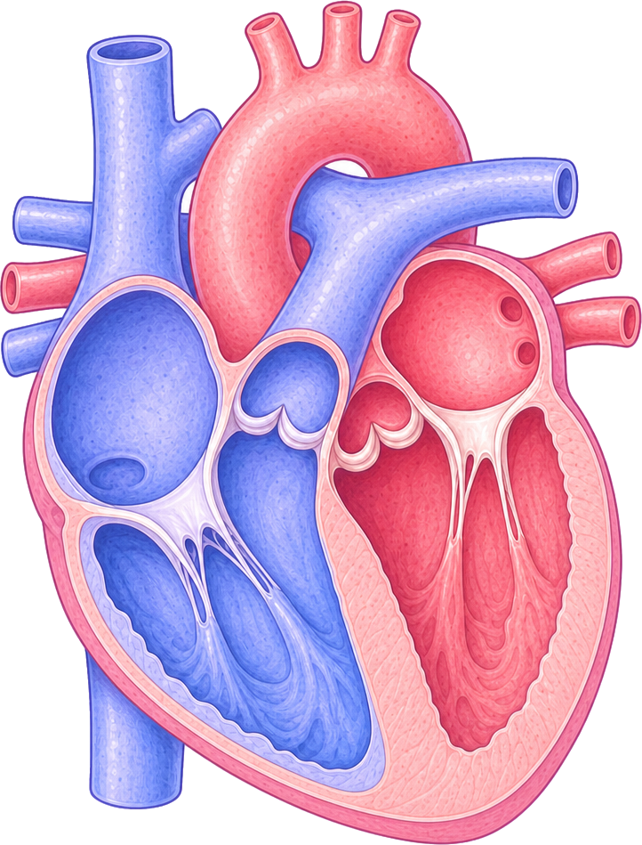 Superior vena cava
Right atrium
Right ventricle
Inferior vena cava
Aorta
Pulmonary artery
Pulmonary veins
Left atrium
Left ventricle
Septum
Heart Diagram
A human heart diagram with the four chambers, aorta, vena cava, pulmonary vessels and septum labeled. Also as a blank version for quizzes.
PNG 1400×900 · HD 2800×1800 · SVG with editable labels
Free for personal, classroom and commercial use. A credit is appreciated, not required. Paste this under it:
<a href="https://sciencedrawings.com/">Drawing by sciencedrawings.com</a>Label the heart
A free labeling quiz: drag each label to the box it belongs in, or tap a label and then tap a box. Great for revision, homework and class.
What each part does
- Superior vena cava
- Brings oxygen-poor blood from the head and arms back into the right atrium.
- Right atrium
- Collects oxygen-poor blood from the body and passes it down to the right ventricle.
- Right ventricle
- Pumps oxygen-poor blood out through the pulmonary artery to the lungs.
- Inferior vena cava
- Brings oxygen-poor blood from the lower body back into the right atrium.
- Aorta
- The largest artery; carries oxygen-rich blood from the left ventricle to the whole body.
- Pulmonary artery
- Carries oxygen-poor blood from the right ventricle to the lungs to pick up oxygen.
- Pulmonary veins
- Bring oxygen-rich blood back from the lungs into the left atrium.
- Left atrium
- Collects oxygen-rich blood from the lungs and passes it down to the left ventricle.
- Left ventricle
- The strongest chamber, with the thickest wall; pumps oxygen-rich blood into the aorta.
- Septum
- The muscle wall down the middle that keeps oxygen-poor and oxygen-rich blood apart.
More like this
 Circulatory System Diagramlabeled + blank
 Brain Diagramlabeled + blank
 Digestion Process Diagramlabeled + blank
 Digestive System Diagramlabeled + blank
All human body drawings · Download the human body pack (ZIP)