 sciencedrawings
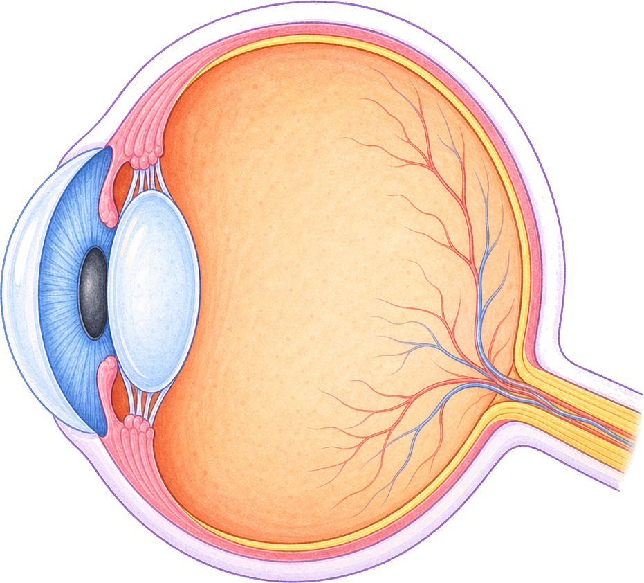 Ciliary muscle
Iris
Pupil
Cornea
Lens
Vitreous humour
Sclera
Retina
Optic nerve
Eye Diagram
A cross-section of the human eye with the cornea, iris, pupil, lens, retina, optic nerve and more labeled. Also as a blank version for quizzes.
PNG 1400×900 · HD 2800×1800 · SVG with editable labels
Free for personal, classroom and commercial use. A credit is appreciated, not required. Paste this under it:
<a href="https://sciencedrawings.com/">Drawing by sciencedrawings.com</a>Label the eye
A free labeling quiz: drag each label to the box it belongs in, or tap a label and then tap a box. Great for revision, homework and class.
What each part does
- Ciliary muscle
- Pulls on the lens to change its shape so the eye can focus near or far.
- Iris
- The coloured ring of muscle that changes the size of the pupil to control how much light gets in.
- Pupil
- The hole in the middle of the iris that lets light into the eye.
- Cornea
- The clear, curved front window of the eye that bends light as it comes in.
- Lens
- Changes shape to focus light onto the retina.
- Vitreous humour
- The clear jelly that fills the eyeball and keeps it round.
- Sclera
- The tough white outer layer that protects the eye and keeps its shape.
- Retina
- The light-sensitive layer at the back of the eye, with rods and cones that detect light.
- Optic nerve
- Carries signals from the retina to the brain.
More like this
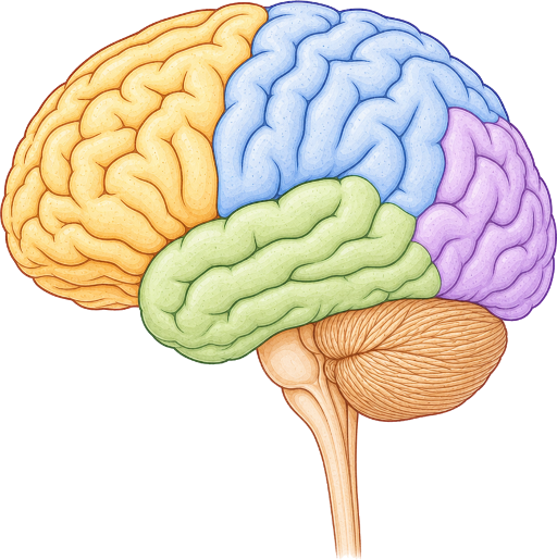 Brain Diagramlabeled + blank
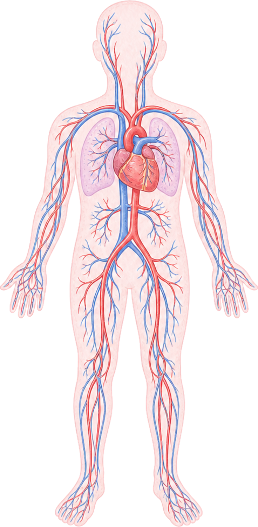 Circulatory System Diagramlabeled + blank
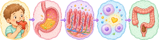 Digestion Process Diagramlabeled + blank
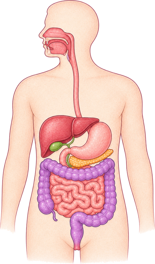 Digestive System Diagramlabeled + blank
All human body drawings · Download the human body pack (ZIP)