 sciencedrawings
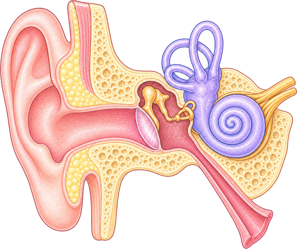 Ossicles
Pinna
Ear canal
Eardrum
Semicircular canals
Auditory nerve
Cochlea
Eustachian tube
Ear Diagram
A labeled ear diagram of the outer, middle and inner ear: pinna, eardrum, ossicles, cochlea and more. Labeled and blank versions.
PNG 1400×900 · HD 2800×1800 · SVG with editable labels
Free for personal, classroom and commercial use. A credit is appreciated, not required. Paste this under it:
<a href="https://sciencedrawings.com/">Drawing by sciencedrawings.com</a>Label the ear
A free labeling quiz: drag each label to the box it belongs in, or tap a label and then tap a box. Great for revision, homework and class.
What each part does
- Ossicles
- Three tiny bones (hammer, anvil and stirrup) that pass the eardrum's vibrations to the inner ear and make them stronger.
- Pinna
- The outer flap of the ear, which collects sound waves and funnels them into the ear canal.
- Ear canal
- The tube that carries sound waves from the outside to the eardrum.
- Eardrum
- A thin membrane that vibrates when sound waves hit it.
- Semicircular canals
- Three fluid-filled loops in the inner ear that sense head movement and help us keep our balance.
- Auditory nerve
- The nerve that carries signals from the cochlea to the brain, where they are heard as sound.
- Cochlea
- A snail-shaped, fluid-filled tube that turns vibrations into nerve signals.
- Eustachian tube
- A tube linking the middle ear to the throat that keeps the air pressure on both sides of the eardrum equal.
More like this
 Brain Diagramlabeled + blank
 Circulatory System Diagramlabeled + blank
 Digestive System Diagramlabeled + blank
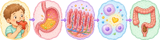 Digestion Process Diagramlabeled + blank
All human body drawings · Download the human body pack (ZIP)